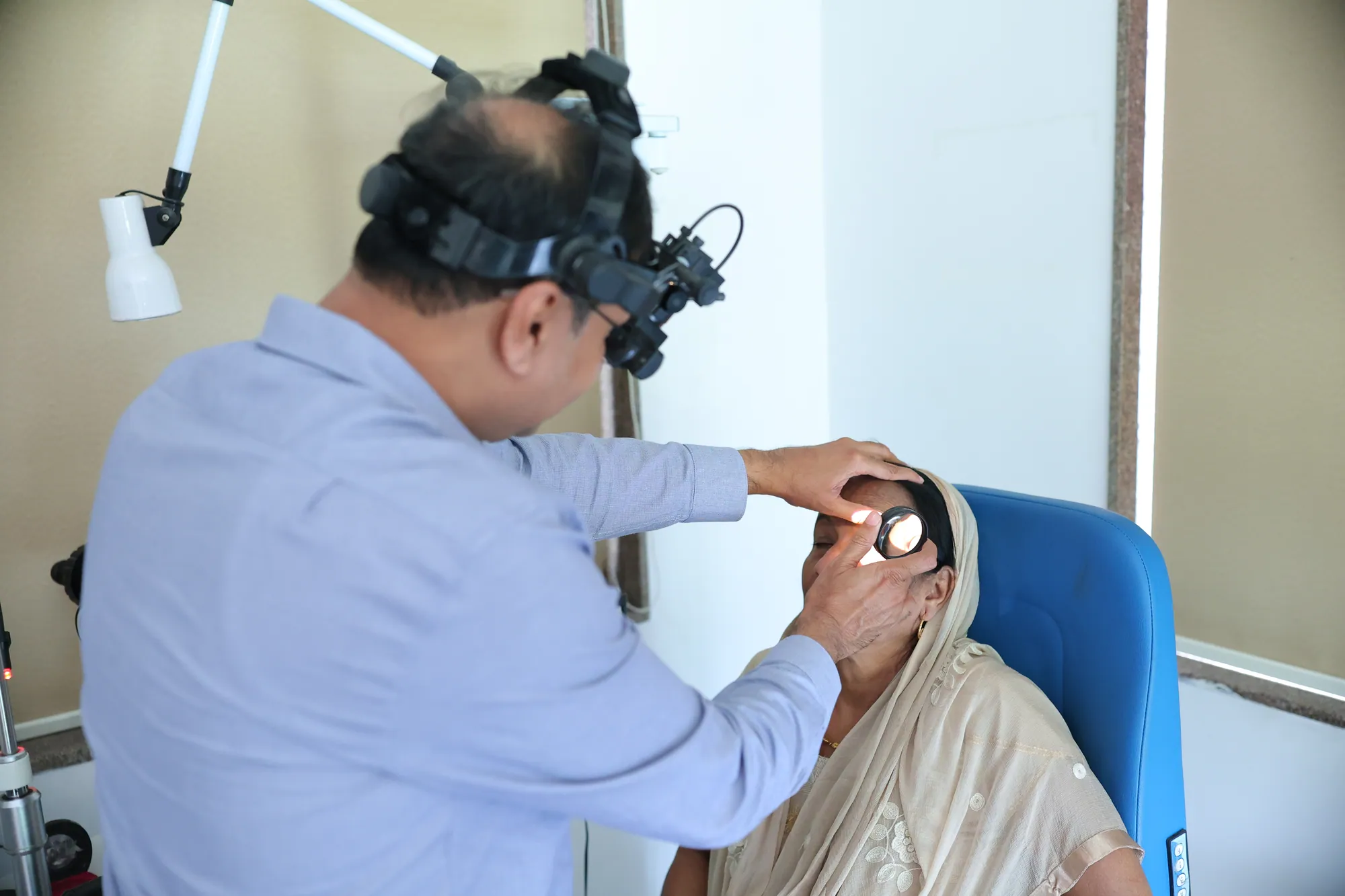
The retina is a light-sensitive layer at the back of the eye. It receives the focused image and sends signals through the optic nerve to the brain. Retinal concerns can affect central detail, side vision, colour or the appearance of spots and flashes. They may be associated with diabetes, age-related changes, inflammation, injury or other eye conditions; only an examination can identify the cause. A retina page is an educational starting point, not a diagnosis, and symptoms that are sudden or severe should not wait for a routine appointment.
Krishna Hospital’s retina pathway brings together a specialist discussion and, when the clinician believes it is useful, appropriate imaging or tests. The aim is to explain what a finding means, whether monitoring or treatment is reasonable, and when referral or another service is needed. Retina care includes medical assessment and, for selected problems, surgical evaluation. Those are not interchangeable labels: the right pathway depends on the diagnosis, examination and available clinical plan. Patients should receive a clear explanation before any test, injection, laser or operation is scheduled.
What the retina does and why it matters
The retina contains specialised cells that respond to light. The macula, near its centre, supports detailed vision used for reading and recognising faces. The peripheral retina contributes to side vision and can be involved in tears or detachment. Because different parts of the retina support different functions, one person may notice distortion or a central blur while another describes flashes, floaters or a shadow. The symptom alone does not identify the cause; a clinician may need a dilated examination or imaging to see the structures clearly.
Retinal conditions are not limited to people who already know they have an eye disease. Long-term health conditions, previous eye procedures, family history, injury and age may be relevant to a clinician’s assessment. Some changes are gradual and can be subtle. Others appear quickly and need prompt review. Mention whether the change is in one or both eyes, when it began, whether it is worsening, and whether it followed an injury or procedure. Do not assume that a painless change is harmless or that a familiar floater has the same cause as a new one.
Symptoms worth discussing promptly
Possible retina-related concerns include new distortion of straight lines, a central blur, difficulty reading small detail, reduced colour vividness, spots, flashes, or a change in peripheral vision. People with diabetes may need eye assessments even when they do not notice symptoms, because some retinal changes may not be obvious to them early on. The appropriate frequency and tests are decided by the treating clinician based on eye findings, medical history and local care guidance.
A sudden shower of new floaters, repeated flashes of light, a dark curtain or shadow across the vision, or sudden loss of vision can be a warning of a retinal tear or detachment. These symptoms need same-day urgent eye assessment where possible; do not wait for a standard appointment or depend on an online enquiry reply. A direct blow, penetrating injury or chemical exposure also needs urgent medical care. If you cannot reach the hospital, seek the nearest emergency service. This page cannot determine whether a symptom is an emergency in your individual case.
How a retina assessment may be planned
The specialist usually begins by listening to the timeline and character of the symptoms, checking medical history and reviewing previous eye records. Visual acuity, pupil response, eye pressure or other examinations may be part of a wider eye assessment. A dilated examination lets the clinician view the back of the eye more clearly when appropriate. Some drops temporarily blur near vision or increase light sensitivity, so ask whether you should arrange transport or bring sunglasses after dilation.
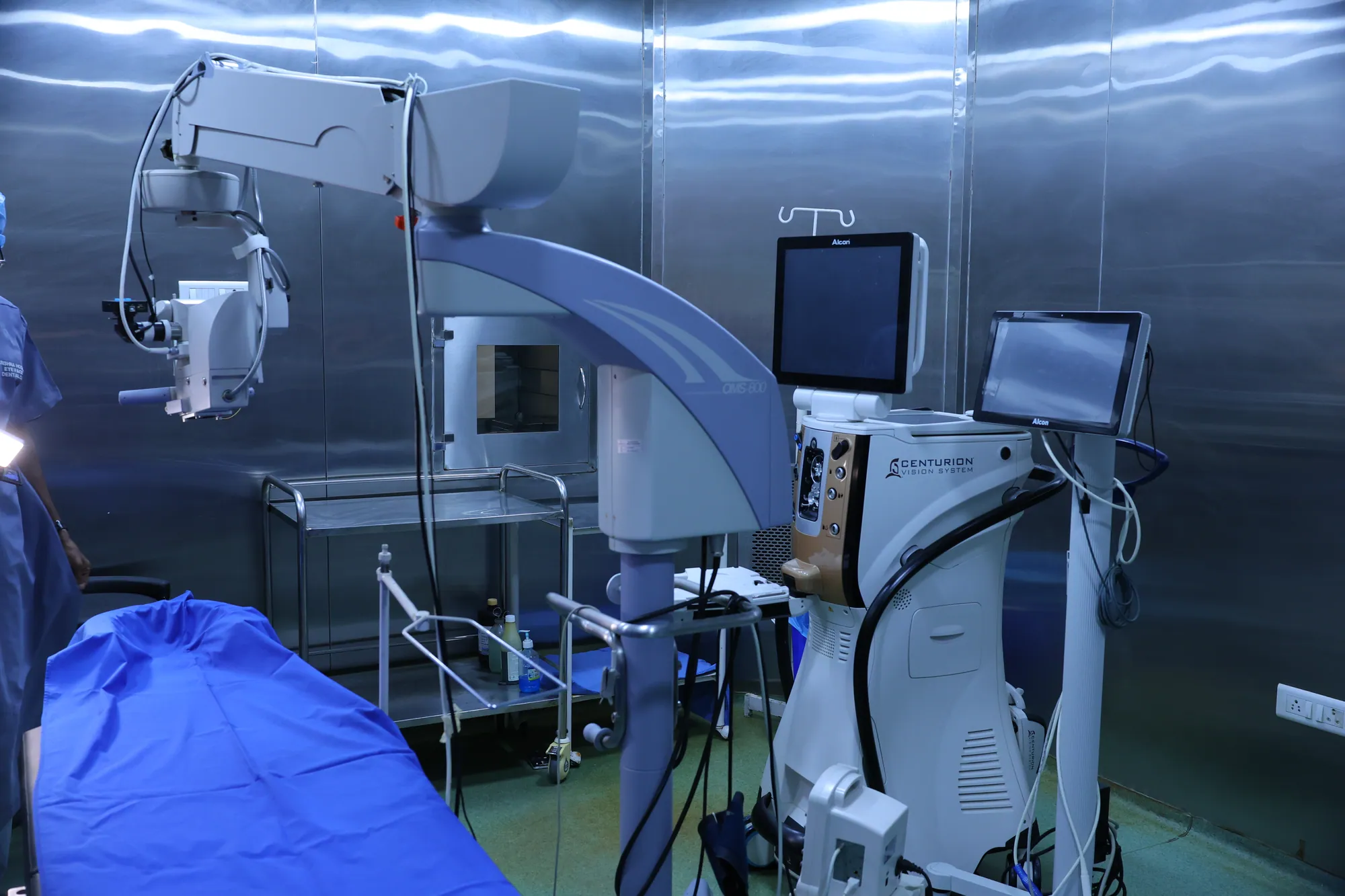
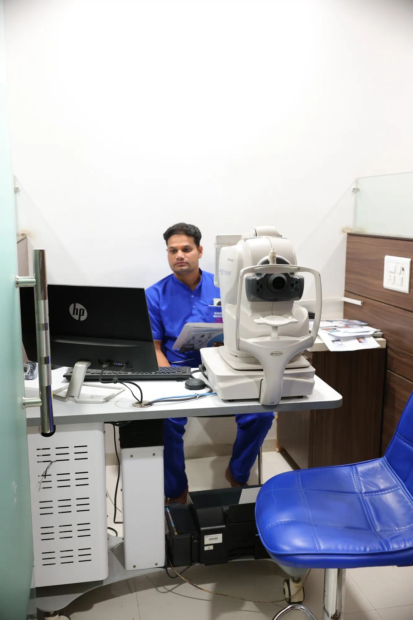
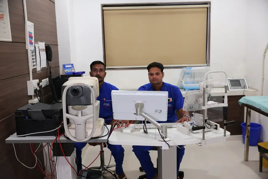
Imaging can help document structures and compare findings over time. The chart supplied by the hospital lists OCT and OCT angiography, as well as fundus photography and green laser. These are different services: an image is not a diagnosis by itself, and the doctor chooses a test based on the question being investigated. The clinician should tell you what a proposed test is intended to show, whether it requires dilation, how long it may take, and whether the fee is separate. A test is not necessary for every patient merely because it appears in a price guide.
Medical retina and surgical retina
Medical retina focuses on evaluating and managing retinal disease through clinical assessment, monitoring and non-surgical options that the clinician considers suitable. Surgical retina is a specialist pathway for selected problems where an operation may be considered after examination. The terms describe different approaches, not a promise that one treatment is needed or available to every person. A retina finding may also require care from another specialty, shared care, a second opinion or referral to a centre with specific services.
For a medical retina discussion, bring relevant diabetes or medical records, earlier eye images and a medication list. For a possible surgical consultation, bring previous operation notes and any imaging already completed. Ask whether the problem is urgent, what the next test is for, what alternatives exist, what the expected follow-up involves and where the procedure would be performed if required. The hospital should confirm the clinician, scope of the appointment and any referral arrangements. No website text can guarantee that a specific surgery will be offered or appropriate.
Understanding common retina tests
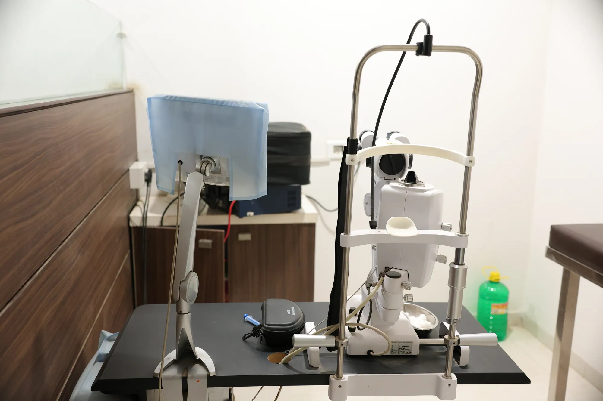
Optical coherence tomography (OCT) produces cross-sectional images of selected eye structures. OCT angiography is a related imaging method that can show blood-flow patterns without being a substitute for a full clinical examination. Fundus photography records a view of the back of the eye and may support documentation or comparison. A retinal laser uses focused light for specific indications chosen by an ophthalmologist. The names sound technical, so it is reasonable to ask the team to explain the purpose and what information is expected before agreeing to a test.
Preparation varies. Some examinations use dilating drops; others may not. A scan can require looking at a fixation target and staying still for a short time. If you have difficulty sitting, focusing, hearing instructions or tolerating bright light, tell the staff before the test. Bring glasses, reports and a companion if helpful. Ask when results will be reviewed and whether they will be discussed in the same visit. The amount printed beside an investigation in the chart does not reveal whether a consultation, repeat images, medicines or follow-up are included.
Making a plan that fits the finding
Recommendations depend on the type, location and stage of a retinal change, the health of the other eye, general health and the person’s needs. A clinician may advise observation with a review schedule, additional imaging, a medical treatment, laser, surgery assessment or referral. Some retinal changes can be monitored; others require faster action. Do not borrow another patient’s plan or delay a review because a symptom seems stable. If a finding or instruction is unclear, ask for it in writing and confirm which symptoms should prompt an earlier call.
A useful care plan states the working explanation in words the patient understands, the next step, the timeframe and who will arrange it. Ask what may happen if a review is missed and whether another clinician needs to know about the result. If cost is a concern, request a written estimate before a planned procedure and ask which charges are included. When a plan involves more than one test, the team can explain the order and whether each one is needed. The patient should have space to ask questions and consider options without pressure.
Living with a retina concern
Keep appointments and bring updated medical information. If diabetes or another chronic condition is part of your history, share the clinician’s advice with the professional coordinating that care. Follow only the instructions given for your own eyes. Do not stop prescribed treatment, use unprescribed drops or try a home remedy to change vision. If sight is reduced, ask the hospital about practical safety steps and support for travel or daily tasks while the assessment is under way.
Use a simple symptom note: the date and time a change began, which eye seems affected, whether it is changing, and what you were doing. This can help explain the timeline, but it does not replace examination. Seek urgent care for sudden vision loss, new flashes with many floaters, a curtain-like shadow, severe eye pain or an injury. If symptoms are stable but worrying, request an assessment and ask when to expect a response. Good retina care is a partnership between examination, clear communication and follow-up—not a single scan or a price on a page.
Keeping records and coordinating care
Retinal care may involve more than one appointment, so keeping reports together can make conversations easier. Store the date of each examination, the name of each test, the clinician’s written explanation and the next review date. If you receive images on a disc, secure link or printed page, bring them to the next visit rather than repeating a test automatically. The specialist can decide whether older images are comparable and still useful. Do not attempt to interpret colour changes or measurements yourself; image quality and clinical context matter.
If another doctor is managing diabetes, blood pressure or a different eye condition, ask whether they need a copy of the retinal report. Share your current medicines and any recent change in health. When a referral is suggested, confirm who sends the records and who is expected to contact you. If you are asked to return within a particular timeframe, write it down and contact the clinic if you cannot attend. An administrative delay should not be used to ignore a sudden symptom; urgent warning signs still need prompt in-person assessment.
Making the discussion understandable
Medical terms such as macula, vitreous, haemorrhage or detachment can sound alarming before their meaning is explained. Ask the clinician to point to a diagram or report and say what it means for your eye. You can ask which part of the finding is certain, what is still being checked, and what change would alter the plan. If you hear a recommendation but do not know why it is needed, repeat it back in your own words and ask the clinician to correct anything you have misunderstood.
It can help to bring someone who can take notes, but the patient should still be included in decisions and consent. Ask about the timing in practical terms: today, within a few days, or at a routine review. Request a phone number or appointment route for a worsening symptom. If you need information in Hindi or another language used by your family, tell the staff so they can try to explain the plan clearly. A scan, printed fee or web page should support—not replace—a two-way conversation with the treating professional.
For a child or an older adult who depends on a caregiver, explain what assistance is available at home and during travel. The clinician can tell you whether that support changes the appointment plan or preparation instructions.